“Targeting Everything from Cancer Growth to Immune Evasion with a Single Gene”... Multitarget Gene Therapy Strategy Proposed for Triple-Negative Breast Cancer
- Seoul National University Hospital: Delivering the TIF1γ gene to triple-negative breast cancer patient tissue reduces cancer cell proliferation and increases apoptosis
- Mouse experiments showed tumor suppression and no discernible lung metastatic lesions; cell experiments showed enhanced phagocytosis of cancer cells by macrophages
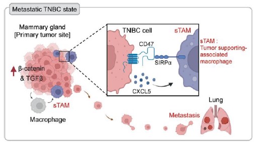
[Figure 1] Metastatic status of triple-negative breast cancer. Within cancer cells, β-catenin and TGFβ signaling is activated, promoting proliferation and migration. At the same time, through CD47 and CXCL5, macrophages (sTAMs) stop attacking the cancer cells and instead support them, and the cancer cells metastasize to the lungs. (sTAM: supporting tumor-associated macrophage)
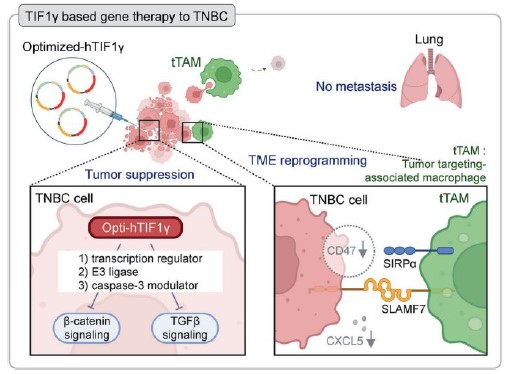
[Figure 2] TIF1γ gene therapy. Delivery of the codon-optimized TIF1γ gene leads to high-level expression of the TIF1γ protein within cancer cells, which inhibits β-catenin, TGFβ, and STAT3 signaling and reduces cancer cell proliferation and migration. At the same time, CD47 and CXCL5 decrease, and macrophages regain the ability to engulf (phagocytose) cancer cells instead of supporting them (tTAMs). By targeting both the cancer cells themselves and the tumor microenvironment, this approach suppresses cancer progression. (tTAM: tumor-targeting tumor-associated macrophage)
In triple-negative breast cancer—characterized by early recurrence and limited targeted therapies—a single gene has been shown to inhibit cancer cell growth and metastasis while also promoting the ability of macrophages to engulf (phagocytose) cancer cells. These effects were confirmed in cultured cells, patient tissue, and mouse models.
On October 8, a research team led by Professor Hyo-Soo Kim of the Biomedical Research Institute and Professor Eun Ju Lee of the Department of Biomedical Science at Seoul National University Hospital (with Dr. Hyomin Park) announced that they had improved the endogenous TIF1γ (transcriptional intermediary factor 1 gamma; a gene naturally present in the body) gene, delivered it to preclinical models of triple-negative breast cancer, and confirmed its multitarget antitumor effects.
The findings were published in the latest issue of Signal Transduction and Targeted Therapy (Impact Factor: 81.2), an international journal published by Springer Nature.
Triple-negative breast cancer is a type of breast cancer defined by the absence of estrogen receptor, progesterone receptor, and human epidermal growth factor receptor 2 (HER2). Accounting for approximately 20% of all breast cancers, it is characterized by early recurrence and a tendency to metastasize (spread) to other organs. Because few targeted therapies are available, treatment relies primarily on chemotherapy. Even existing targeted therapies have limitations: because they block only a single signaling pathway, cancer cells can activate compensatory (bypass) signaling routes to escape the blockade.
The research team focused on TIF1γ, a protein that regulates various cellular signals; its expression is frequently reduced in cancer. The team first showed in 2020 that TIF1γ inhibits liver fibrosis and, in 2022, developed a “smart gene therapy” technology that activates the gene only in diseased liver. They extended its application to pulmonary fibrosis in 2025 and have now expanded it to cancer. This study used a “codon-optimized” gene (opti-hTIF1γ), whose nucleotide sequence was modified so that cells produce more protein.
The team cultured tumor tissues from seven patients with triple-negative breast cancer at Seoul National University Hospital ex vivo (outside the body) and delivered this gene into them. In cancer cells that received the gene, vimentin (a protein associated with metastatic potential) and Ki67 (a marker of proliferation) decreased markedly, while apoptosis (programmed cell death) increased. Both markers also decreased in patient cancer cells that had spread outward from the tissue. When the same experiment was performed on mouse tumor tissue, the number of cancer cells spreading outward from the tissue fell by approximately 70%.
The mouse experiments also showed clear differences. Human triple-negative breast cancer cells were implanted into the mammary fat pad of mice, and 14 days later, lipid capsules (liposomes) carrying the gene were injected around the tumors. At the end of the experiment, tumors in the control group had grown to more than 300 mm³, whereas tumors in the treatment group were significantly smaller or hardly observable. The area occupied by proliferating cancer cells (Ki67-positive area) was also reduced to less than 10% of that in the control group.
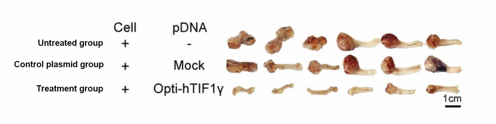
[Figure 3] Mouse tumors. Tumors in the control group grew to more than 300 mm³, whereas those in the treatment group were significantly smaller or hardly observable.
Metastasis also differed. Injected cancer cells were detected in the lymph nodes of the control group but not in the treatment group. In the control group, the alveolar structure of the lungs was severely disrupted by cancerous lesions, whereas the lungs of the treatment group maintained normal structure. In another metastasis model—the mammary intraductal (MIND) model, in which cancer cells are injected directly into the mammary ducts—cancer cells pretransfected with the gene did not give rise to discernible lung metastatic lesions.
Unlike conventional methods that block only a single signal, TIF1γ showed a “multitarget” effect by simultaneously inhibiting three key signaling pathways involved in cancer progression. In cultured triple-negative breast cancer cells, TIF1γ first induced degradation of β-catenin, a protein that drives cancer growth and migration; second, suppressed TGFβ signaling, which promotes metastasis; and third, induced degradation of the STAT3 protein, which supports cancer cell survival and immune evasion.
The immune environment also changed. In patient-derived cancer cells that received the gene, CD47—an immune-evasion protein that cancer cells display to avoid macrophage attack—and CXCL5—a molecule involved in recruiting tumor-supporting macrophages—decreased together. The research team suggests that STAT3 inhibition was involved in these changes.
Macrophage responses changed as well. In macrophages co-cultured with cancer cells showing reduced CD47 and CXCL5, SLAMF7, a receptor that promotes phagocytosis, increased. In co-culture of cancer cell lines and macrophages, cancer cells that received the gene were engulfed by macrophages or died, and in patient tissue cultures, fragments of cancer cells were observed inside macrophages. The team named these macrophages, which regained the ability to engulf cancer cells, “tumor-targeting macrophages (tTAMs).”
Because this was a preclinical study, the team plans follow-up research, such as delivery technologies that can also be used for widely disseminated (systemically spread) cancer, to enable application in actual patients.
Professor Hyo-Soo Kim (Cardiovascular Research Group) said, “While conventional targeted therapies were limited to blocking a single signal, this study confirmed that TIF1γ alone can simultaneously inhibit multiple signals involved in cancer cell growth and metastasis and restore the phagocytic ability of macrophages.” He added, “This shows the potential of multitarget gene therapy that addresses both the cancer cells themselves and the tumor microenvironment.”

[From left] Professors Hyo-Soo Kim (Biomedical Research Institute), Eun-Ju Lee (Department of Biomedical Science) and Dr. Hyo-Min Park, Seoul National University Hospital