"Spatial Map" of Esophageal Cancer Tissue Created After Chemoradiotherapy… Radiation Resistance-Linked ΔNp63/NRF2 Gene Set Highest at "Tumor Leading Edge"
- Seoul National University Hospital and Seoul Metropolitan Government-Seoul National University Boramae Medical Center analyzed post-treatment surgical tissues from 56 patients, identifying eight distinct spatial regions.
- Among patients with regression grades 1–2, higher scores for inflammation-related gene sets in the "Fibro-Immune Niche" were associated with an increased risk of disease progression, recurrence, and death.
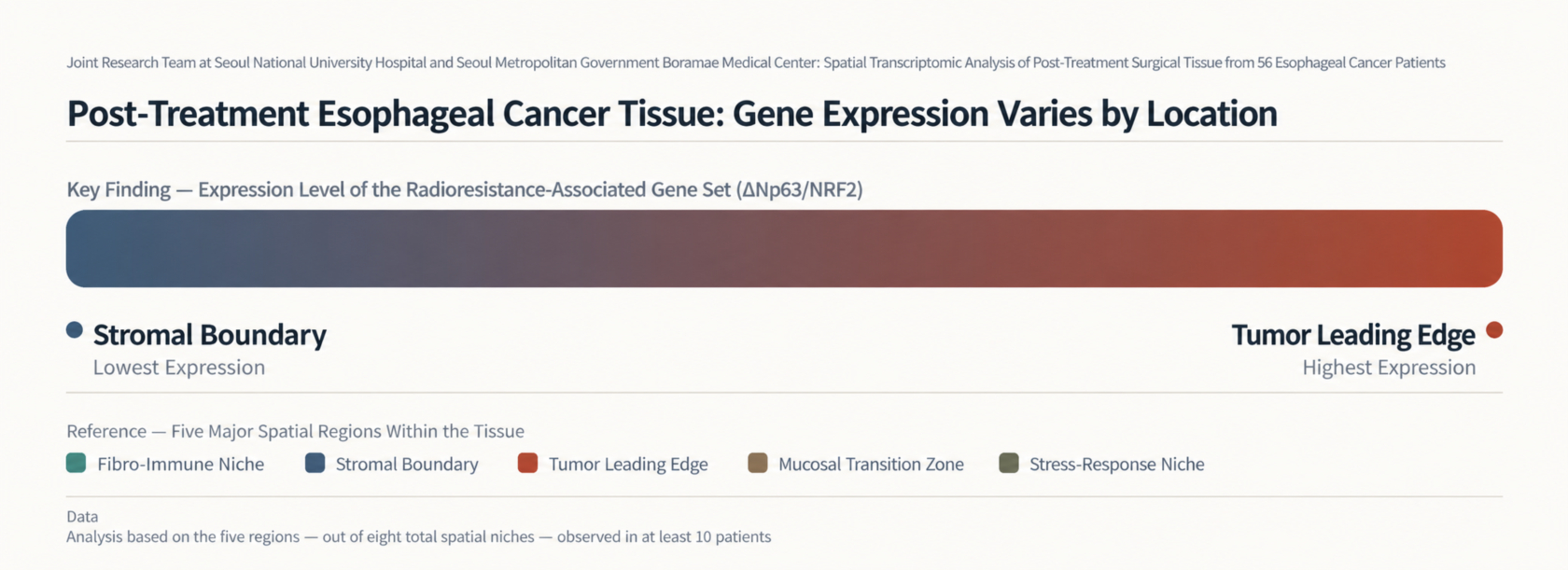
[Figure 1] Expression of the ΔNp63/NRF2 gene family (implicated in radioresistance) was highest at the "Tumor Leading Edge" and lowest at the "Stromal Boundary."
Following chemoradiotherapy, esophageal cancer tissues exhibited distinct gene expression patterns depending on their specific location within the tumor. A domestic research team analyzed surgical tissue samples from 56 patients by region and found that the ΔNp63/NRF2 gene cluster—previously linked to radiation resistance—showed the highest expression levels at the "Tumor Leading Edge" and the lowest at the "Stromal Boundary." In an exploratory analysis of 27 patients whose clinical outcomes were difficult to predict based solely on pathological grading, higher scores for inflammation-related gene clusters in the "Fibro-Immune Niche" were associated with an increased risk of disease progression, recurrence, and death.
On October 1, a joint research team—comprising Professors Bum-Sup, Jang and Hak-Jae, Kim (Department of Radiation Oncology), Kwon-Joong, Na (Department of Thoracic and Cardiovascular Surgery), and Hye-Seung, Le (Department of Pathology) at Seoul National University Hospital, along with former Professor Byoung-Hyuck, Kim (Department of Radiation Oncology at Seoul Metropolitan Government-Seoul National University Boramae Medical Center)—announced these findings. The team reached this conclusion after analyzing stored surgical tissue samples from 56 patients with esophageal squamous cell carcinoma who underwent esophagectomy following preoperative chemoradiotherapy between 2005 and 2022.
Esophageal squamous cell carcinoma is a cancer that arises from the squamous cells of the esophageal mucosa. Some patients with locally advanced disease—where the cancer has spread to surrounding tissues or lymph nodes but has not metastasized to distant organs—undergo chemoradiotherapy prior to surgery. Although many patients still have residual tumors following treatment, the spatial characteristics of this remaining tissue have not been fully elucidated.
Conventional single-cell analysis involves dissociating tissue for measurement, making it difficult to determine the specific location within the tissue where gene expression occurs. The research team utilized spatial transcriptomics technology (Visium), which allows for the measurement of gene expression while preserving spatial information.
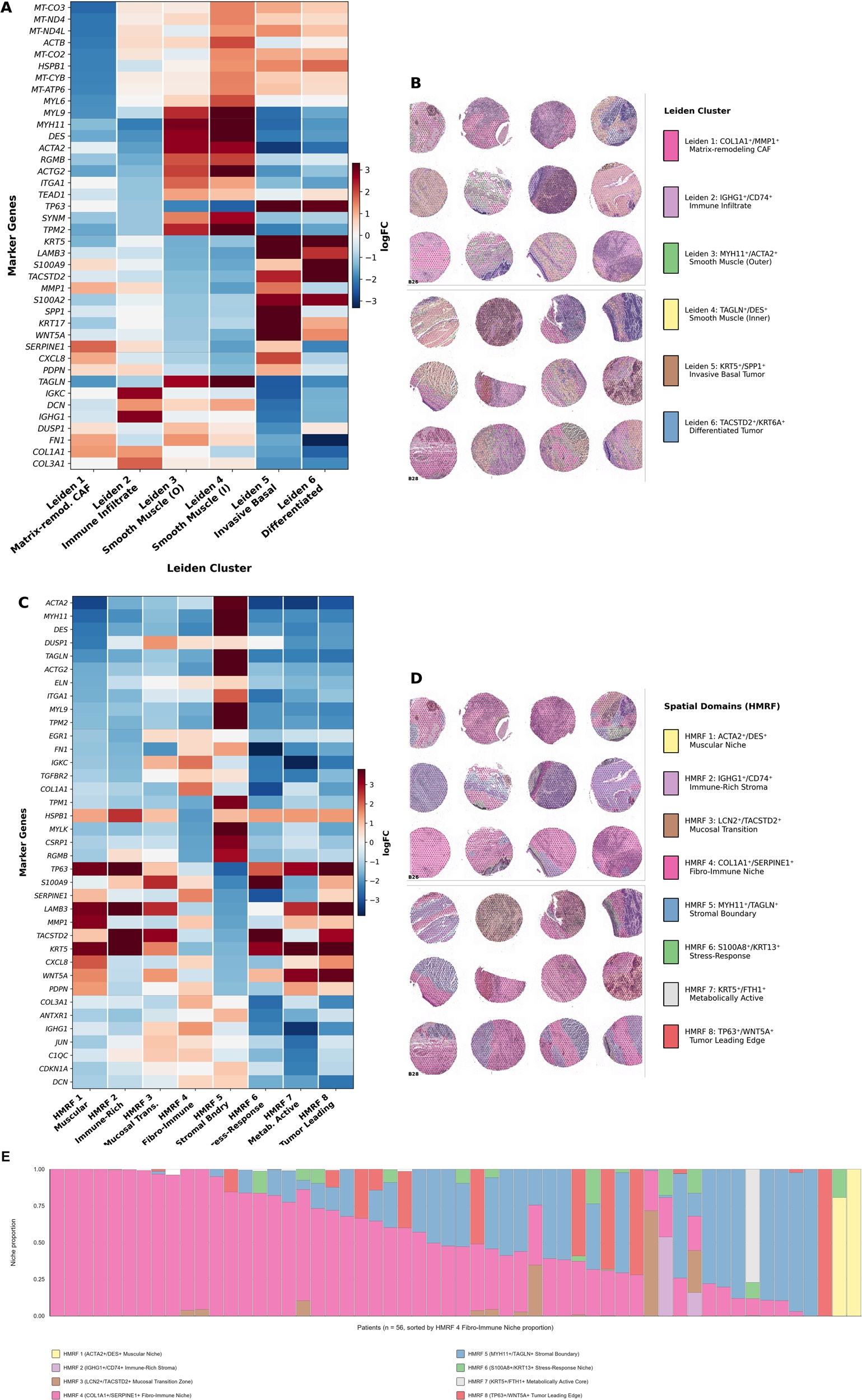
[Figure 2] Spatial transcriptomic map of esophageal cancer surgical specimens following chemoradiotherapy. A and B show gene expression clusters; C and D show eight spatial regions; and E displays the proportion of these regions for each of the 56 patients.
The research team analyzed tissue microarrays obtained from the surgical tissues of 56 patients and categorized the tissue into eight spatial regions. Among these, five major regions—observed in at least 10 patients—were identified: the Tumor Leading Edge, the Fibro-Immune Niche, the Stromal Boundary, the Mucosal Transition Zone, and the Stress-Response Niche. The Fibro-Immune Niche was the largest, accounting for an average of 53.0% of the patient tissue.
When the research team compared the expression scores of 37 gene sets across these five major regions, all 37 sets showed statistically significant differences depending on the region. Notably, the ΔNp63/NRF2 gene set showed the highest expression levels in the Tumor Leading Edge and the lowest in the Stromal Boundary.
The research team also analyzed whether these spatial characteristics correlated with the patients' actual clinical outcomes. Among the pathological regression grades—an indicator, scored from 0 to 3, of how much of the tumor disappeared in the surgical tissue—they focused on 29 patients classified as Grade 1 or 2 (indicating residual cancer), categories where long-term prognosis is difficult to predict based on standard pathological regression grading alone, and specifically examined the 27 individuals among them who exhibited the Fibro-Immune Niche.
The results showed that for five specific gene sets associated with the Fibro-Immune Niche—inflammation, ΔNp63/NRF2, SPP1, multigene radiotherapy-resistance, and oncogenic transcription factors—higher scores tended to correlate with a shorter "failure-free survival" period (the time elapsed without disease progression, recurrence, or death). For every one-standard-deviation increase in the gene set score, the risk of progression, recurrence, or death rose by 1.76 to 2.53 times; the strongest association was observed with the inflammation-related gene set, showing a 2.53-fold increase in risk.
A similar trend was observed across the entire patient cohort. Among the 52 patients evaluated for the Fibro-Immune Niche, higher scores for the gene set associated with SPP1, ΔNp63, and NRF2 correlated with poorer outcomes—specifically regarding overall survival, failure-free survival, and survival free from local recurrence or distant metastasis—with a 1.36- to 1.62-fold higher risk. However, the research team noted that as this was an initial exploratory study using post-treatment tissue samples, these findings serve as a candidate for further validation in future research.
Professor Bum-Sup Jang (Department of Radiation Oncology, Seoul National University Hospital) stated, "This study established a spatial map of esophageal cancer tissue following chemoradiotherapy and revealed that tumors and surrounding tissues exhibit distinct molecular characteristics based on their location." He added, "It will serve as a meaningful foundation for understanding the post-treatment tumor microenvironment and advancing future precision medicine research."
The findings of this study were published in the latest online edition of the international journal npj Precision Oncology (Impact Factor: 9.9).
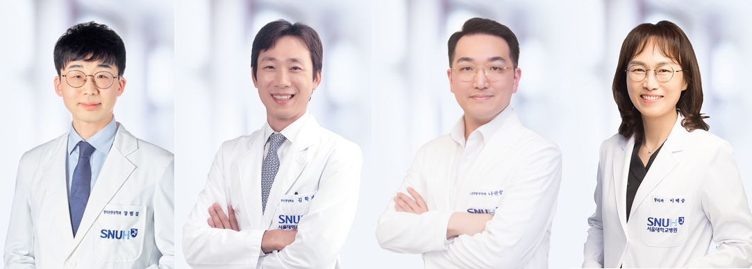
[From left] Professors Bum-Sup, Jang and Hak-Jae, Kim (Department of Radiation Oncology), Professor Kwon-Joong, Na (Department of Thoracic and Cardiovascular Surgery), and Professor Hye-Seung, Lee (Department of Pathology) at Seoul National University Hospital.