SNUH Research Team Develops AI Model for Dementia Screening and Risk Stratification Using Retinal Fundus Photographs
- Joint research team from SNUH, SNU College of Medicine, and XAIMED Co., Ltd., analyzes 108,008 retinal fundus images from routine health checkups
- The AI model achieved an AUROC of 0.750 for dementia detection (screening) and a C-index of 0.812 for predicting future dementia incidence (risk stratification)
A domestic research team in South Korea has developed an artificial intelligence (AI) model capable of screening for current dementia status and predicting future dementia risk using only retinal fundus photographs. Crucially, by applying ‘Explainable AI’ techniques, the model visualizes the specific retinal regions that influenced its predictions, thereby enhancing its feasibility for clinical translation.
Dementia is difficult to diagnose using a single definitive test and typically requires a comprehensive evaluation combining clinical assessments, cognitive testing, and brain magnetic resonance imaging (MRI). However, cost and accessibility limitations hinder the widespread use of these modalities for population-level screening. Conversely, the retina is considered an accessible extension of the central nervous system that mirrors cerebral vascular and neuronal changes, making retinal fundus photographs a promising, noninvasive biomarker for dementia risk assessment.
A joint research team—led by Professor Park Sang-min (Department of Family Medicine, SNUH), Dr. Chang Joo-young (XAIMED Co., Ltd.), Research Assistant Professor Han Chang-ho (Medical Big Data Research Center, SNU College of Medicine), and Dr. Kim Jae-won (Department of Biomedical Sciences, SNU College of Medicine)—developed and evaluated a deep learning framework for dementia screening and prediction using 108,008 retinal fundus images from 36,322 individuals who underwent routine health checkups at SNUH between 2004 and 2016.
The research team defined 1,001 individuals (2,868 images) diagnosed with dementia prior to or within two years of fundus examination as dementia-positive cases, matching them 1:4 with non-dementia controls to construct a model development dataset. Using this dataset, they evaluated 15 distinct AI models created by combining five vision foundation models with three fine-tuning strategies.
After comparing the predictive performance across all models to select the optimal pipeline, the team evaluated its clinical utility and explainability. Explainability was validated by generating saliency heatmaps using Vision Transformer attention rollout to visualize the regions influencing AI decisions, followed by quantitative comparison against retinal anatomical structures, including the optic disc and retinal vessels.
The results demonstrated that the AI model utilizing the ‘RETFound-MAE’ foundation model—pre-trained on millions of retinal images—combined with a partial fine-tuning strategy yielded the highest performance. This optimal model achieved an AUROC of 0.750 for current dementia detection and a C-index of 0.812 for predicting future dementia incidence, significantly outperforming the conventional clinical dementia risk scoring tool, CAIDE (AUROC 0.624, C-index 0.689).
Even after adjusting for established dementia risk factors such as age and sex, higher AI output scores remained significantly associated with increased odds and hazards for current and future dementia. This demonstrates that retinal fundus images contain incremental information relevant to dementia risk beyond traditional clinical risk factors.
In decision curve analysis evaluating clinical utility, the AI model provided a higher net benefit than the CAIDE score, screen-all strategy, and screen-none strategy. Furthermore, combining the AI output with individual CAIDE component variables improved the predictive performance (AUROC) from 0.749 to 0.774, confirming that the AI model can complement existing clinical risk-stratification frameworks.
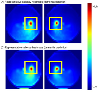
[Image] Saliency heatmaps illustrating the retinal regions prioritized by the artificial intelligence model. Warmer red colors indicate areas of stronger model focus. For both dementia detection (A) and future incidence prediction (B), the highest attention was consistently identified in the optic disc and adjacent peripapillary regions.
Analysis of the model’s decision-making rationale revealed high attention focused on the ‘optic disc’ and ‘peripapillary areas.’ The relative saliency intensity in the optic disc was 5.72 times higher than in reference non-vessel, non-optic disc regions, with the highest attention observed specifically in the temporal quadrant of the optic disc. High saliency was also confirmed in the papillomacular bundle connecting the optic disc and macula, as well as the peripapillary region. These findings align closely with previously reported dementia-related retinal structural changes in medical literature, supporting the conclusion that the AI relies on biologically plausible retinal signals.
Professor Park Sang-min (Department of Family Medicine, SNUH) stated, “We hope this AI technology, which utilizes routine health checkup data without requiring additional high-cost diagnostic procedures, will serve as a valuable complementary tool in dementia prevention and early intervention strategies.”
Dr. Chang Joo-young (XAIMED Co., Ltd.) commented, “This study presents an important direction for developing fundus image-based AI biomarkers by systematically evaluating not only predictive performance but also explainability and clinical utility.”
This study was published in the latest issue of the international academic journal ‘npj Digital Medicine (Impact Factor 18.0).’
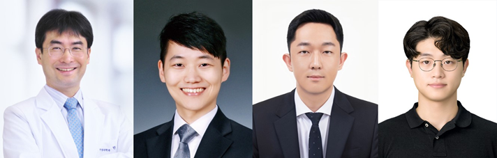
[Photo from left] Professor Park Sang-min (Department of Family Medicine, SNUH), Dr. Chang Joo-young (XAIMED Co., Ltd.), Research Assistant Professor Han Chang-ho (Medical Big Data Research Center, SNU College of Medicine), and Dr. Kim Jae-won (Department of Biomedical Sciences, SNU College of Medicine).