Seoul National University Hospital First Identifies Molecular Biological Characteristics of ‘VETC-type Liver Cancer’ with Poor Prognosis
- Confirmation of Unique Characteristics of VETC Liver Cancer via Spatial Transcriptome Analysis… Validation of a 276-Case Cohort
- Identification of ‘LAMTOR2’ Gene, Prognosis Prediction, and Suggestion of Potential Therapeutic Target
Among hepatocellular carcinomas, there is a type characterized by particularly frequent recurrence and a poor prognosis. This occurs when the tumor mass is completely surrounded by blood vessels, exhibiting a "vessels encapsulating tumor clusters (VETC)" pattern. A research team at Seoul National University Hospital has confirmed that the LAMTOR2 gene is prominently present in this type of liver cancer.
A research team led by Professor Hye-Ryoung Kim of the Department of Pathology, Professor Seok-Kyun Hong of the Department of Hepatobiliary and Pancreatic Surgery at Seoul National University Hospital, and Professor Hyun-Je Kim of the Department of Medical Science at Seoul National University (along with Professor Yun-Jung Hwang of the Department of Pathology at Seoul National University Bundang Hospital and Researcher San-Ha Hwang) announced on the August 20th that they identified the unique molecular and metabolic characteristics of VETC liver cancer by analyzing liver cancer surgical tissue using spatial transcriptome techniques.
Liver cancer was the second leading cause of cancer death in Korea in 2024, following lung cancer, with hepatocellular carcinoma accounting for the majority of primary liver cancers. According to statistics from the National Center for Caner Knowledge and Information for 2019–2023, the 5-year relative survival rate is only 40.4%; furthermore, the presence of a VETC pattern indicates a high risk of recurrence after surgery and a lower survival rate.
While the VETC pattern serves as a prognostic indicator that can be identified relatively easily through tissue biopsy alone, it has not been elucidated which specific molecular signals define VETC-positive hepatocellular carcinoma. This is because conventional bulk RNA sequencing has struggled to capture subtle differences based on location within the tumor.
Consequently, the research team focused on the 'spatial transcriptome' technique, which examines gene expression while preserving locational information within the tissue. Through this method, they aimed to identify new biomarkers representing VETC patterns by confirming molecular differences between VETC-positive and negative regions.
To this end, tissue samples were collected from patients who underwent surgery for hepatocellular carcinoma at Seoul National University Hospital from September 2022 to March 2023. For spatial transcriptome analysis, 42 regions were utilized, including 18 tumor liver tissue samples (7 VETC-positive and 11 VETC-negative) and 24 non-tumor liver tissue samples. For verification, 276 cases of immunohistochemistry (IHC) and 5 cases of single-cell RNA sequencing were also examined.
The analysis results showed that compared to the VETC-negative regions, the number of genes increased by 39 and decreased by 235 at the VETC-positive regions. The increased genes were closely related to angiogenesis and fatty acid metabolism pathways, while conversely, immune-related pathways were suppressed.
In particular, four genes (LAMTOR2, DPP4, ZNHIT1, and MLXIPL) were consistently higher than in VETC-negative tissue and normal liver; among them, only LAMTOR2 was selected as a key candidate because it was found to be tumor-specifically increased in The Cancer Genome Atlas (TCGA). LAMTOR2 is a gene that activates mTOR signaling, which regulates cell growth and metabolism.
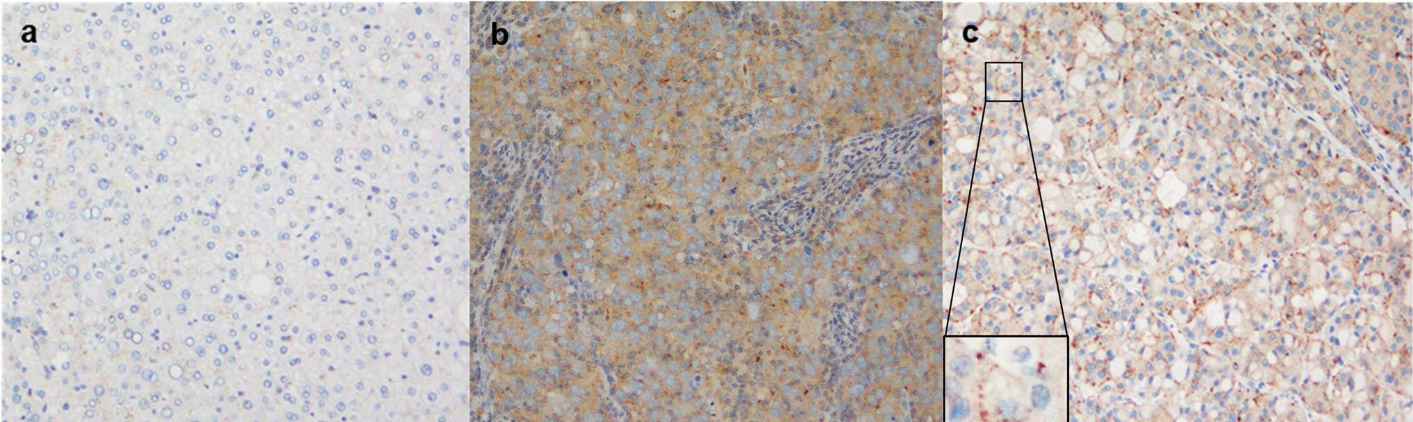
[Figure] Representative immunohistochemical (IHC) images of LAMTOR2 expression: a. Normal liver tissue b. VETC-negative tumor cells c. VETC-positive tumor cells ('peripheral accentuation' pattern with darker staining edges observed)
Immunohistochemical testing was performed to verify whether the narrowed LAMTOR2 had actually increased at the protein level. As a result, a 'peripheral accentuation' pattern was observed, in which the margins of tumor cells were heavily stained with LAMTOR2. A distinct difference was observed, with 71% (5/7) in VETC-positive liver cancer and 27% (3/11) in VETC-negative liver cancer.
When verified with a cohort of 276 cases, the patient group exhibiting the peripheral accentuation pattern had a VETC positivity rate of 37%, which was higher than in cases without the accentuation pattern (20%). The recurrence rate after surgery was also 75%, exceeding that of the control group (57%), suggesting that the peripheral accentuation pattern is linked to prognosis.
The same trend was confirmed at the single-cell level. LAMTOR2 appeared prominently in cell groups with high VETC association among malignant hepatocytes. In these cell groups, metabolic pathways related to lipid degradation and detoxification (cytochrome P450) were also activated.
Professor Hye-Ryoung Kim (Department of Pathology) stated, “We expect that the molecular biological characteristics of VETC-positive liver cancer identified in this study will serve as important foundational data for the future development of personalized treatment methods.”
Professor Hyun-Je Kim (Department of Medical Science, Seoul National University) explained, “This study started with spatial transcriptome analysis and was cross-validated step-by-step using public databases, large-scale tissue cohorts, and single-cell analysis,” adding, “This multi-faceted verification underpins the reliability of the discovery of LAMTOR2.”
Professor Seok-Kyun Hong (Department of Hepatobiliary and Pancreatic Surgery) said, “This study is significant in that it identified the hidden molecular characteristics of VETC-positive hepatocellular carcinoma, which has a poor prognosis, through spatial transcriptome analysis,” and added, “We will continue follow-up research to ensure that LAMTOR2 can evolve into a target for personalized treatment and a prognosis prediction indicator.”
Meanwhile, the results of this study were published in the online edition of the international academic journal ‘Hepatology International’.

[Photo from left] Professor Yun-Jung Hwang(Department of Pathology, Seoul National University Bundang Hospital), Researcher San-Ha Hwang(Seoul National University Cancer Research Institute), Professor Hye-Ryoung Kim(Department of Pathology, Seoul National University Hospital); Professor Seok-Kyun Hong(Department of Hepatobiliary and Pancreatic Surgery), Professor Hyun-Je Kim(Department of Medical Science, Seoul National University)