Seoul National University Hospital Achieves World's First Recellularization of Xenogeneic Pericardial Valve Scaffolds Using a Single Stem Cell Monoculture
- Xenoantigens causing immune rejection removed, followed by single-cell stem cell monoculture… tissue regeneration and calcification-inhibiting effects confirmed
- Simplifies previous co-culture method requiring two cell types… paves the way for next-generation bioprosthetic heart valves
A research team at Seoul National University Hospital (SNUH) has succeeded for the first time in the world in recellularizing xenogeneic heart valve tissue using a single type of stem cell — reviving decellularized animal-derived scaffolds into living-tissue-like constructs — while reducing the risk of immune rejection and calcification, two chronic problems associated with animal-derived valves used in cardiac surgery. The achievement opens a path toward new treatment options for patients with congenital heart disease, who have historically faced repeated valve replacement surgeries.
The research team, led by Professor Hong-Gook Lim of the Department of Pediatric Thoracic & Cardiovascular Surgery at SNUH (with Research Professor So-Young Kim of the Biomedical Research Institute and Professor Gi-Beom Kim of the Department of Pediatric Cardiology), announced on July 27 that it had confirmed both in vitro and in vivo recellularization, along with calcification-inhibiting effects, by seeding stem cells alone onto xenoantigen-removed porcine pericardial scaffolds.
Porcine and bovine pericardial and valve tissues are currently widely used as graft materials in cardiac surgery. However, these tissues retain xenoantigens not found in humans, which trigger immune rejection and, over time, lead to calcification — a major cause of graft failure. A new technology was therefore needed both to remove these xenoantigens and to enable the valve to become stably integrated within the body.
The team had previously succeeded in vitro in recellularizing enzymatically xenoantigen-removed porcine pericardium by co-culturing human adipose-derived mesenchymal stem cells (hADSCs) with human umbilical vein endothelial cells (HUVECs). Because this co-culture approach required simplification for clinical translation, the team conducted a follow-up study establishing a more practical single-cell monoculture recellularization strategy using hADSCs alone.
The team decellularized porcine pericardial tissue and treated it with two enzymes (α-galactosidase and PNGase-F) in combination to remove the xenoantigens α-Gal and Neu5Gc. The scaffold surface was then coated with a fibrin mesh (with heparin and VEGF) to promote cell attachment, producing a xenoantigen-free pericardial scaffold. Human adipose-derived stem cells were seeded onto this scaffold and cultured for 8 weeks to observe in vitro recellularization. Separately, scaffolds that underwent decellularization, glutaraldehyde (GA) fixation, and anti-calcification treatment were seeded with rat bone marrow-derived mesenchymal stem cells (BM-MSCs) and implanted subcutaneously in rats to evaluate in vivo recellularization and the degree of calcific deposition.
![[그림1] 체외 재세포화 실험 (영문)](https://www.snuh.org/upload/editor/images/000115/20260803155426668_FJML7CDF.jpg "external_image")
[Figure 1] In vitro recellularization: After 56 days of hADSC monoculture on porcine pericardial scaffolds treated with both α-galactosidase and PNGase-F, recellularization was enhanced, with increased expression of associated marker proteins (green/red).
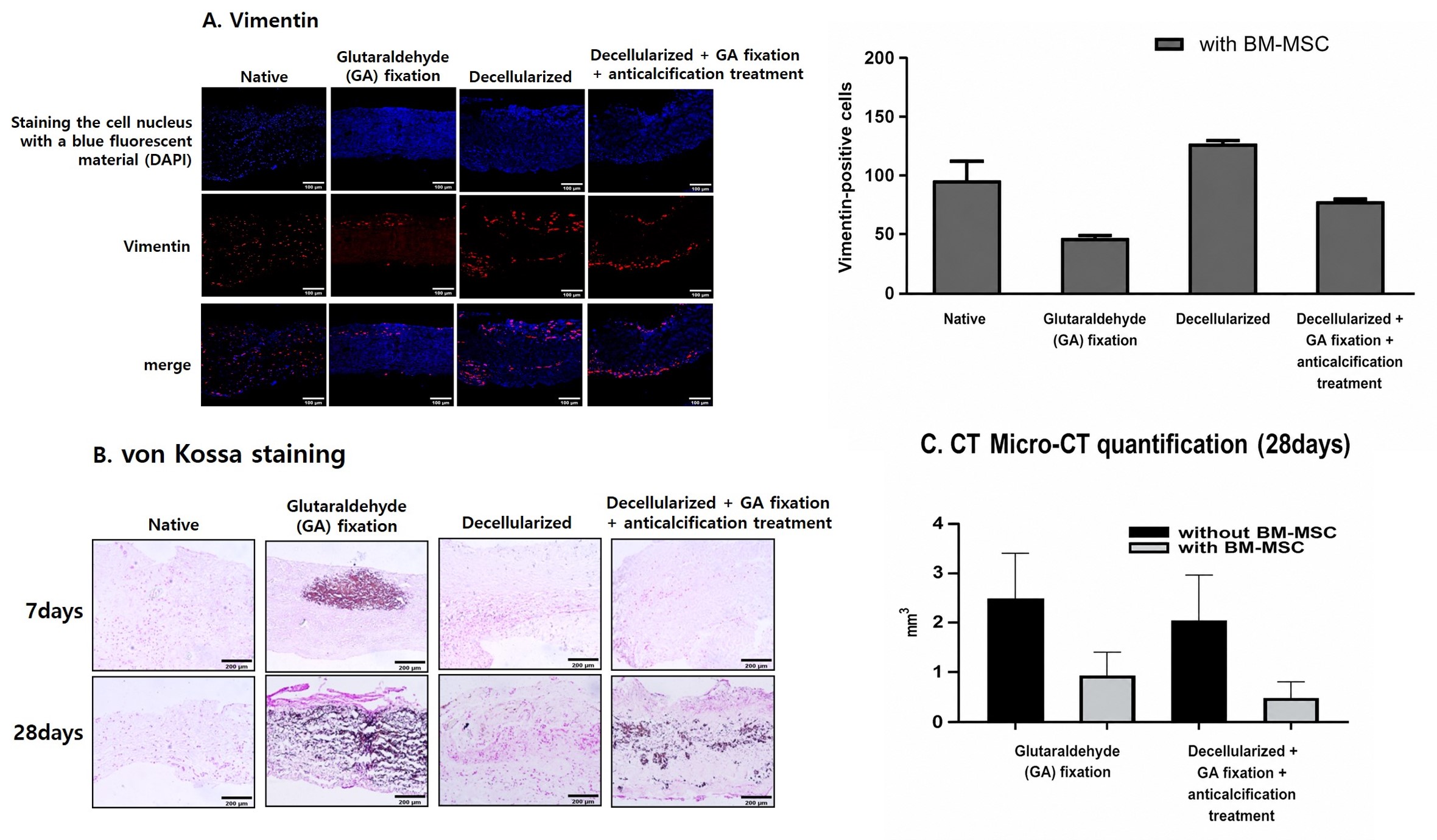
[Figure 2] In vivo experiment: In scaffolds recellularized after decellularization, GA fixation, and anti-calcification treatment: (A) increased vimentin expression (red) on histological analysis; (B) reduced calcific deposits (dark staining) on von Kossa staining; (C) significantly reduced calcium deposition on quantitative micro-CT analysis.
Results showed that scaffold tissue architecture was preserved when both enzymes were used following decellularization. Seeded stem cells had infiltrated into the tissue matrix by day 28, and by day 56 cells were evenly distributed across the surface and throughout the interior, with cell infiltration and tissue remodeling becoming progressively more pronounced over time.
Expression of recellularization-associated proteins — vimentin, fibronectin, calponin, CD31, and von Willebrand factor (vWF), together with phalloidin staining of the cytoskeleton — all increased. This indicates that the stem cells did not merely attach to the scaffold surface but infiltrated the tissue, differentiated into new cell types, and drove recellularization.
In the animal experiments, the implanted scaffolds also became stably integrated. Among grafts recellularized after decellularization, GA fixation, and anti-calcification treatment, vimentin expression was more pronounced, and calcification levels were significantly lower than in non-recellularized grafts.
Professor Hong-Gook Lim (Department of Pediatric Thoracic & Cardiovascular Surger) said, "This study is significant in that it integrates the removal of immunological barriers, cell engraftment, tissue remodeling, and calcification inhibition into a single platform," adding, "We will continue this research so that the technology can eventually be translated into patient treatment."
This study was supported by the Kun-Hee Lee Child Cancer & Rare Disease Project, and the findings were published in the online edition of Bioengineering, an international journal in the field of biomedical engineering.
Separately, the research team previously applied its own anti-calcification protocol to porcine pericardium to develop the PULSTA™ transcatheter pulmonary valve in 2018, which has since been implanted in 1,075 patients with excellent clinical outcomes. In January 2026, the technology also passed CE MDR (Medical Device Regulation) Class III certification in Europe, meeting rigorous safety and efficacy standards and expanding its clinical use globally, including in the European market.
![[사진 왼쪽부터] 의생명연구원 김소영 연구교수, 소아청소년과 김기범 교수, 소아흉부외과 임홍국 교수](https://www.snuh.org/upload/editor/images/000115/20260803155426751_9JWSUNQY.jpg "external_image")
[Photo from left] Research Professor So-Young Kim (Biomedical Research Institute), Professor Gi-Beom Kim (Department of Pediatric Cardiology), Professor Hong-Gook Lim (Department of Pediatric Thoracic & Cardiovascular Surgery)