Seoul National University Hospital Develops Noninvasive IV-MRL to Visualize Lymphatic Vessels via Simple Intravenous Injection
- "Lymphatic phase" identified 20–30 minutes post- contrast medium injection, with gadolinium concentration in lymph surpassing that in blood
- Successfully visualizes hepatic and mesenteric lymphatics, overcoming limitations of conventional diagnostic imaging
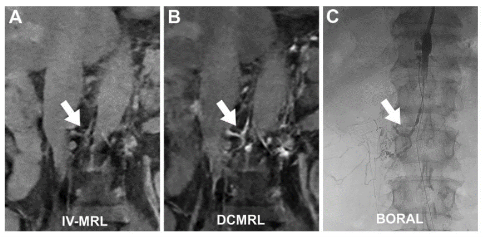
[Figure 1] Comparative analysis of the newly developed IV-MRL against conventional methods (DCMRL, BORAL). Unlike traditional diagnostic procedures (B, C) that mandate invasive lymph node punctures or catheter placements, IV-MRL approach (A) seamlessly visualizes the comprehensive lymphatic system via an effortless intravenous contrast injection.
A South Korean research team has developed a pioneering noninvasive MRI technique, Intravenous Contrast-Enhanced MR Lymphangiography (IV-MRL), which visualizes the lymphatic system through a standard peripheral intravenous injection instead of a technically demanding direct injection into the inguinal lymph nodes. Seoul National University Hospital (SNUH) announced on the Julyt 9th that the team successfully visualized the hepatic and mesenteric lymphatics, structures that were previously difficult to evaluate using conventional imaging modalities.
The lymphatic system plays a critical role in maintaining immune function and fluid balance. However, when lymphatic vessels are damaged—often as a result of oncologic surgeries—it can lead to lymphatic leakage, causing severe complications such as nutritional deficiencies, compromised immunity, and chylous ascites. Consequently, a rapid and accurate diagnostic tool is essential to localize the exact sites of leakage.
Currently, inguinal intranodal lymphangiography and Dynamic Contrast-Enhanced MR Lymphangiography (DCMRL) serve as the standard diagnostic methods for the conducting lymphatics. However, these approaches require precise needle placement into the inguinal lymph nodes, making them technically challenging. Furthermore, they rarely visualize the hepatic and mesenteric lymphatics, which are responsible for generating over 70% of total body lymph. This structural limitation has hindered accurate diagnosis, restricting the leakage detection rate to approximately 55% in patients with chylous ascites, where lymph fluid accumulates in the peritoneal cavity due to mesenteric lymphatic injury.
To address these challenges, a joint research team led by Professors Sae-Beom Hur (Department of Radiology, SNUH), Sung-Hwan Yoon (Biomedical Research Institute, SNUH), and Lyo-Min Kwon (Department of Radiology, Hallym University Medical Center) evaluated the feasibility of the newly developed noninvasive IV-MRL technique through animal models and a pilot clinical study.
In the swine model experiment, the researchers administered intravenous gadolinium-based contrast agents (Gd-DOTA and Gd-EOB-DTPA) and tracked concentrations in the blood and lymph over time. The study revealed a distinct "lymphatic phase" occurring 20 to 30 minutes post-injection, during which the gadolinium concentration in the lymph exceeded those in the portal and systemic veins. On MR imaging, the signal intensity (SI) ratio of the cisterna chyli* and thoracic duct increased by up to approximately 5.5-fold compared to the surrounding muscle tissue, providing distinct and clear visualization of the lymphatic pathways.
*Cisterna chyli : Sac-like tissue located below the thoracic lymphatic vessels
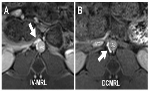
[Figure 2] Visual comparison between IV-MRL and DCMRL. IV-MRL (A) successfully visualizes the hepatic and mesenteric lymphatic structures, which are typically unidentifiable or poorly rendered on conventional DCMRL (B) scans.
When comparing IV-MRL with the conventional DCMRL method, both techniques successfully highlighted the thoracic duct and retroperitoneal lymphatics. However, the intravenous approach demonstrated its capability to visualize hepatic and mesenteric lymphatic structures, which remained poorly visualized on conventional DCMRL scans.
The pilot human study further substantiated these findings. The research team retrospectively analyzed data from 16 patients (mean age: 56.6 years) clinically suspected of lymphatic leakage who underwent IV-MRL between September 2024 and August 2025. While the initial post-contrast imaging at 1 minute yielded a thoracic duct-to-inferior vena cava SI ratio of just 0.49, this ratio substantially increased to 1.47 at the 20-minute mark, demonstrating statistical significance (P < .001).
These findings indicate that IV-MRL can effectively highlight the central conducting lymphatics through a simple peripheral IV line, while extending diagnostic visualization to the elusive hepatic and mesenteric networks.
Professor Sae-Beom Hur (Department of Radiology at SNUH) said "Unlike conventional intranodal techniques, IV-MRL utilizes a standard peripheral vein injection administered by a nurse. This greatly simplifies the procedural workflow and significantly reduces the patient's pain burden. We expect this technique to substantially improve the diagnostic accuracy for lymphatic leakages and aid in establishing targeted treatment strategies."
This study was published in the July issue of Radiology (Impact Factor: 17.6), an international peer-reviewed journal in the field of medical imaging.

[Photo from left] Professor Sae-Beom Hur (Department of Radiology, SNUH), Professor Sung-Hwan Yoon (Biomedical Research Institute, SNUH), and Professor Lyo-Min Kwon (Department of Radiology, Hallym University Medical Center).
