Reducing Unnecessary Biopsies After Kidney Transplant... Blood Test Proven for ‘Precise Rejection Screening’
- Seoul National University Hospital Identifies Non-Invasive Risk Assessment Strategy Combining ‘Donor-Specific Antibodies and Cell-Free DNA’
- Diagnostic Performance (AUC) Improved When Combining Two Tests... 97.8% Probability of No Rejection in Low-Risk Groups with Low Values
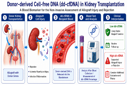
[Figure] Schematic diagram of the principle for non-invasively assessing the risk of rejection by measuring ‘cell-free DNA’ increased in the blood due to transplant kidney damage after kidney transplantation.
The korean research team has demonstrated, through a multi-center prospective study, a "non-invasive risk assessment strategy" capable of minimizing unnecessary invasive biopsies by precisely identifying high- and low-risk groups for asymptomatic rejection after kidney transplantation.
A team led by Professors Ara Jo and Sang-Il Min of the Department of Transplant and Vascular Surgery at Seoul National University Hospital (co-authored with Professor Ju-han Lee of Severance Hospital and Professor Cheol-woong Jeong of Korea University Anam Hospital) announced on the 4th June that performing a "donor-derived cell-free DNA" blood test on patients with newly developed "donor-specific antibodies" after kidney transplantation is useful for predicting asymptomatic rejection.
"Donor-specific antibodies (dnDSA)," newly generated in the body after kidney transplantation, are immunological risk signals and are a major cause of kidney function decline and rejection. However, among patients with these antibodies, the rate at which rejection is confirmed by actual biopsy is only 30–40%. Since the biopsy to confirm this is an invasive procedure involving bleeding, pain, and the burden of hospitalization, a significant number of patients had to endure unnecessary procedural burdens.
As an alternative, donor-derived cell-free DNA (dd-cfDNA) has recently emerged as a non-invasive biomarker for identifying kidney transplant damage. Cell-free DNA refers to "fragments of DNA derived from the transplanted kidney" that are released into the bloodstream when immunological damage or inflammation occurs in the transplanted kidney. However, until now, its clinical utility in determining whether to perform a biopsy on these patients has been uncertain.
The research team prospectively compared blood cell-free DNA levels with biopsy results in 123 patients (77 in the donor-specific antibody positive group and 46 in the negative group) who maintained stable kidney function after kidney transplantation at three transplant centers in Korea.
The analysis revealed that the median blood cell-free DNA level was significantly higher in the antibody-positive group (1.2%) than in the negative group (0.3%). Furthermore, the DNA levels showed a characteristic where they increased proportionally with the severity of microvascular inflammation within the kidney. In fact, when applying the international biopsy diagnostic criteria (Banff Index), the median cell-free DNA level in patients with almost no inflammation in the renal microvessels (inflammation score 0–1) remained at 0.54%, but in patients with severe inflammation (inflammation score 2 or higher), it was found to be higher than 1.6%.
Diagnostic accuracy has also improved. When predicting rejection based solely on the presence or absence of specific antibodies, as was done previously, the overall diagnostic performance indicator (AUC) was 0.74; however, when the two tests were combined and analyzed, the diagnostic performance significantly increased to 0.81.
In particular, this combined testing method is significant in that it reduces unnecessary invasive biopsies, thereby minimizing the procedural burden on patients. Previously, when specific antibodies were detected, the majority of patients had to undergo a biopsy for confirmation, even though the actual probability of rejection was only half (46.2%). However, by adding a cell-free DNA test to identify patients with levels below 1.0%, the probability that they would not experience actual rejection (negative predictive value) reached 97.8%. In other words, combining the two blood tests has established clinical grounds for effectively identifying the majority of low-risk groups and safely deferring biopsies.
Professor Sang-il Min (Department of Transplant Vascular Surgery of Seoul National University Hospital) emphasized, “When donor-specific antibodies are detected after a kidney transplant, both patients and medical staff worry about rejection; however, performing tissue biopsies on all patients in the same manner can be burdensome.” He added, “This study demonstrated that by combining non-invasive biomarkers, it is possible to more precisely identify patients with a high actual risk of rejection.”
He further stated, “If cell-free DNA is appropriately integrated into the clinical decision-making process in the future, it could evolve into a personalized monitoring strategy that detects transplant damage early while reducing the burden on patients.”
The results of this study were published in the April online edition of the international academic journal ‘International Journal of Surgery (IF: 10.3).’

[From left in the photo] Professors Sang-il Min and Ara Jo of the Department of Transplant Vascular Surgery at Seoul National University Hospital
